When Is Vitrectomy Recommended?
Vitrectomy is recommended for patients with retinal or vitreous conditions that cannot be effectively treated with medications or laser therapy. The procedure helps restore vision, repair retinal damage, and prevent permanent vision loss.
- Retinal Detachment: To reattach the retina and preserve vision.
- Vitreous Hemorrhage: To remove blood from the vitreous cavity caused by diabetes, trauma, or retinal diseases.
- Macular Hole & Epiretinal Membrane: To repair macular disorders that cause blurred or distorted central vision.
- Retinal Tears: To treat retinal tears and prevent progression to retinal detachment.
- Diabetic Retinopathy: For advanced diabetic eye disease with persistent bleeding or scar tissue formation.
- Eye Trauma: To remove foreign bodies, repair retinal injuries, and restore the internal structures of the eye.
What Happens During Vitrectomy Surgery?
Before vitrectomy, your retina specialist performs a comprehensive eye evaluation using advanced diagnostic tests such as retinal examination, OCT, fundus photography, and other imaging studies to determine the extent of retinal damage and plan the most appropriate treatment.
Vitrectomy Procedure
- Anesthesia: Local or general anesthesia is administered to ensure a comfortable and pain-free procedure.
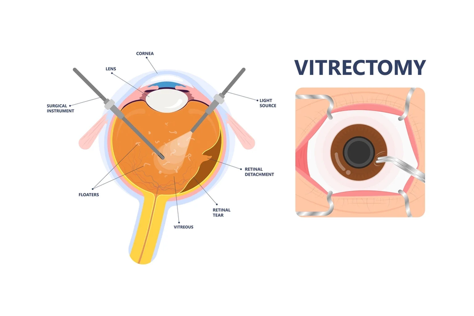
- Small Incisions: Tiny incisions are made in the white part of the eye (sclera) to insert specialized microsurgical instruments.
- Removal of the Vitreous Gel: The cloudy or damaged vitreous gel, along with blood, scar tissue, or foreign material, is carefully removed.
- Retinal Repair: The surgeon repairs retinal tears, macular holes, or retinal detachment using laser treatment or other advanced surgical techniques.
- Replacement of the Vitreous: The eye may be filled with a sterile saline solution, gas bubble, or silicone oil to help support the retina during healing.
- Completion of Surgery: The incisions are closed if required, and a protective eye patch is placed before you are moved to the recovery area.
How to Prepare for Vitrectomy?
Proper preparation helps ensure a safe surgery and smooth recovery. Your retina specialist will provide personalized instructions based on your eye condition, medical history, and overall health.
- Complete Pre-Surgical Evaluation: Undergo the recommended eye examinations, scans, and general health assessments before surgery.
- Follow Fasting Instructions: If advised, avoid eating or drinking for several hours before the procedure, especially if general anesthesia is planned.
- Review Your Medications: Inform your doctor about all medications, including blood thinners, diabetes medicines, and supplements. Take only the medicines approved by your surgeon.
- Arrange Transportation: Plan for someone to accompany you, as you will not be able to drive immediately after surgery.
- Arrive Early: Reach the hospital before your scheduled procedure to complete the necessary formalities and pre-operative preparations.
- Wear Comfortable Clothing: Avoid eye makeup, jewelry, and contact lenses on the day of surgery, and wear loose, comfortable clothing.
Risks & Possible Complications of Vitrectomy
Vitrectomy is a safe and effective procedure when performed by experienced retina specialists. However, like any eye surgery, it carries a small risk of complications that will be discussed with you before treatment.
- Eye Infection: A rare but serious complication that requires immediate medical attention.
- Bleeding Inside the Eye: Minor bleeding may occur during or after surgery and is usually manageable.
- Increased Eye Pressure: Temporary elevation of eye pressure may occur and can be controlled with medication.
- Retinal Detachment: Although uncommon, additional retinal treatment may occasionally be required.
- Cataract Formation: Some patients may develop cataracts more rapidly after vitrectomy, particularly older adults.
- Need for Additional Surgery: In complex retinal conditions, further treatment may be required to achieve the best outcome.
What to Expect After Vitrectomy?
Most patients return home on the same day of surgery. Mild redness, watering, blurred vision, and eye discomfort are common during the first few days and gradually improve as the eye heals.
- Follow Your Medication Schedule: Use prescribed eye drops and medications exactly as directed.
- Protect Your Eye: Wear the protective eye shield and avoid rubbing or putting pressure on the operated eye.
- Maintain Head Position: If a gas bubble is placed inside the eye, follow the specific head positioning advised by your surgeon.
- Avoid Strenuous Activities: Refrain from heavy lifting, bending, swimming, and vigorous exercise until your doctor approves.
- Attend Follow-up Visits: Regular follow-up examinations are essential to monitor healing and retinal recovery.
- Report Warning Signs: Contact your retina specialist immediately if you experience severe pain, increasing redness, sudden vision loss, or persistent flashes and floaters.
Benefits of Vitrectomy Surgery
Vitrectomy is an advanced retinal procedure that helps preserve vision by treating conditions affecting the vitreous and retina. It can significantly improve visual outcomes and prevent permanent vision loss.
- Restores Clear Vision: Removes blood, scar tissue, or cloudy vitreous gel that blocks light from reaching the retina.
- Repairs Retinal Damage: Enables the surgeon to treat retinal tears, macular holes, and retinal detachment with precision.
- Prevents Vision Loss: Helps preserve eyesight in advanced retinal diseases such as diabetic retinopathy and vitreous hemorrhage.
- Improves Visual Function: Restores the normal structure of the retina, improving vision and overall quality of life.
- Treats Complex Eye Conditions: Provides an effective solution for retinal disorders that cannot be managed with medication or laser treatment alone.
Types of Vitrectomy Surgery
The type of vitrectomy performed depends on the location and severity of the eye condition. Your retina specialist will recommend the most suitable approach for your treatment.
Posterior (Pars Plana) Vitrectomy
This is the most commonly performed type of vitrectomy. It involves removing the vitreous gel through tiny incisions in the pars plana to treat retinal detachment, diabetic retinopathy, vitreous hemorrhage, macular holes, and other disorders affecting the back of the eye.
Anterior Vitrectomy
Anterior vitrectomy is performed when vitreous gel moves into the front part of the eye, usually following eye trauma, cataract surgery, or lens-related complications. The procedure helps restore normal eye anatomy and reduces the risk of further complications.
Recovery & Results of Vitrectomy
Recovery after vitrectomy typically takes 2 to 6 weeks, depending on the underlying eye condition and the type of procedure performed. Following your retina specialist’s instructions is essential for proper healing and achieving the best visual outcome.
- Protect the Eye: Wear the recommended eye shield or patch as advised to protect the eye during the initial healing period.
- Use Medications Regularly: Apply prescribed eye drops and medications exactly as directed to reduce the risk of infection and inflammation.
- Follow Positioning Instructions: If a gas bubble is placed inside the eye, maintain the recommended head position to support retinal healing.
- Avoid Strenuous Activities: Refrain from heavy lifting, bending, swimming, and driving until your doctor confirms it is safe.
- Attend Follow-up Visits: Regular post-operative check-ups allow your retina specialist to monitor healing and recovery.
- Expect Gradual Vision Improvement: Vision usually improves over time, although recovery depends on the severity of the retinal condition being treated.