
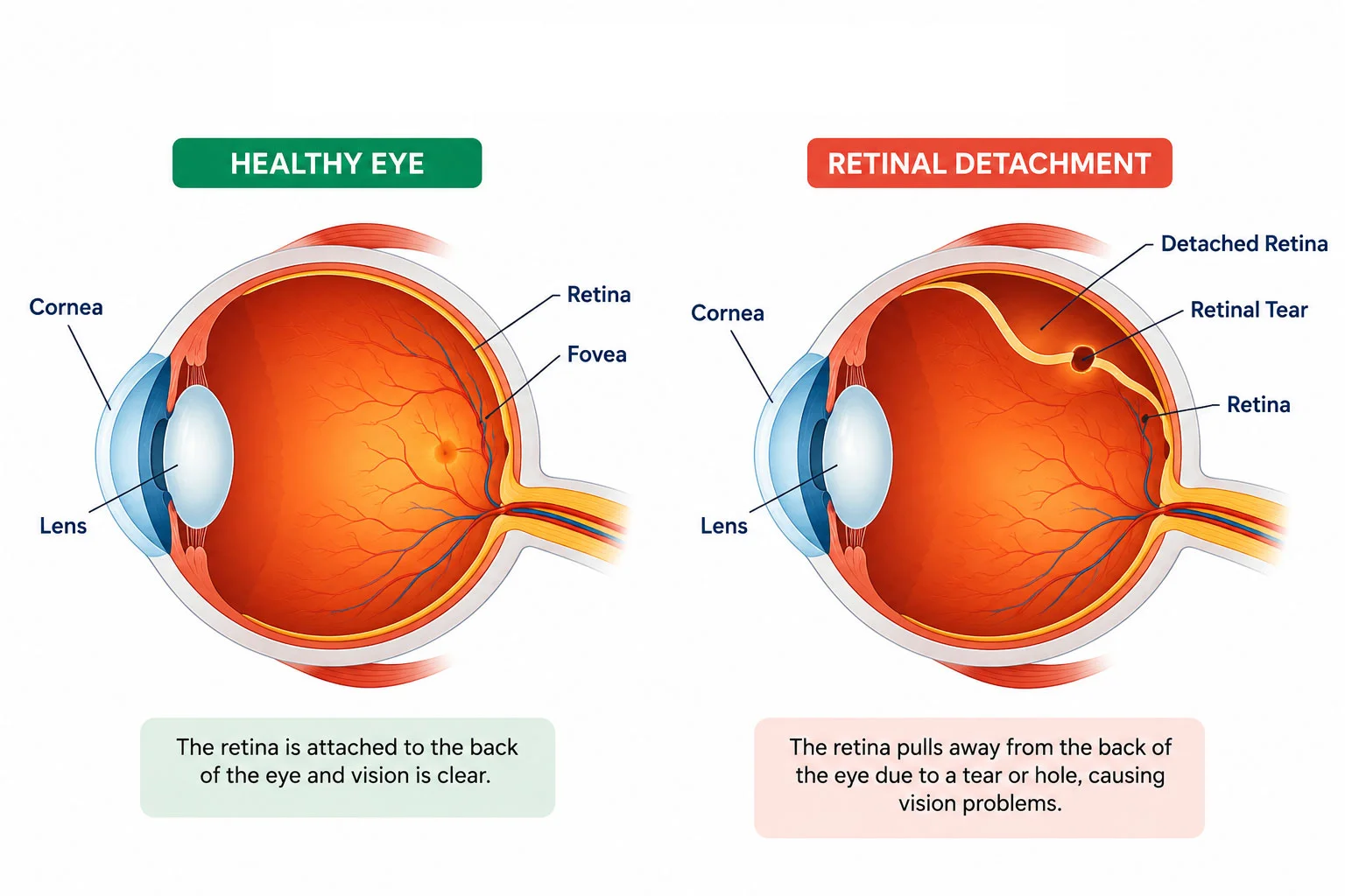
Retinal detachment can occur in different forms depending on its underlying cause. Identifying the type of detachment helps retina specialists determine the most appropriate treatment plan.
The most common type of retinal detachment, caused by a tear or hole in the retina that allows fluid to pass underneath, separating it from the underlying tissue.
Occurs when scar tissue on the retinal surface pulls the retina away from the back of the eye. It is commonly associated with advanced diabetic retinopathy.
Develops when fluid collects beneath the retina without any tear or hole, often due to inflammation, tumors, or certain vascular eye conditions.
Early recognition of retinal detachment symptoms is essential, as prompt treatment can significantly reduce the risk of permanent vision loss.
If you experience any of these warning signs, seek immediate evaluation by a retina specialist. Early diagnosis and timely treatment offer the best chance of preserving vision.
Retinal detachment can occur due to age-related changes or underlying eye conditions. Understanding the risk factors can help with early detection and timely treatment.
Retinal detachment is an eye emergency. Prompt evaluation and treatment can help preserve vision and prevent permanent vision loss.
A detailed retinal examination helps confirm retinal detachment and determine the most appropriate treatment plan.
Eye drops enlarge the pupil, allowing the retina to be examined in detail.
OCT provides detailed cross-sectional images of the retina for accurate assessment.
Ultrasound imaging is used when the retina cannot be clearly seen because of bleeding or cataracts.
High-resolution retinal images help document abnormalities and monitor treatment.
Treatment depends on the type, location, and severity of the retinal detachment. Early intervention offers the best chance of preserving vision.
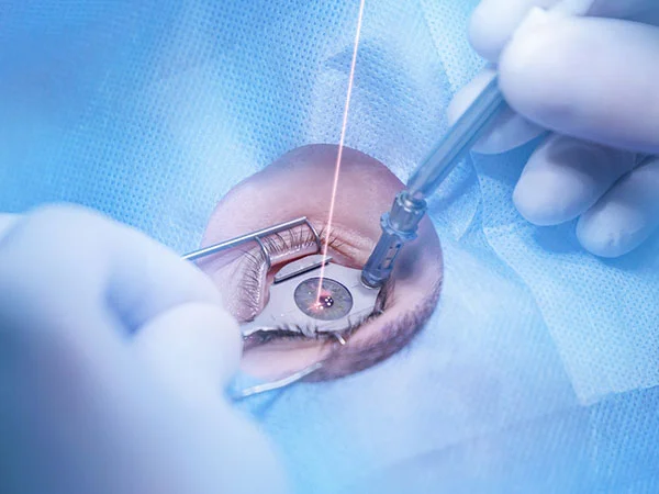
Laser treatment seals small retinal tears before detachment develops.
A freezing treatment used to seal retinal tears and prevent progression.
A gas bubble is injected into the eye to reposition the retina while the tear is sealed.
A silicone band supports the eye and relieves traction on the retina.
The vitreous gel is removed, the retina is repaired, and a gas bubble or silicone oil is used to support healing.
Your retina specialist will recommend the most suitable surgical approach based on your eye condition.
Surgery is performed under local or general anesthesia using advanced microsurgical techniques.
Recovery varies depending on the severity of the detachment and the surgical technique used.
| Recovery Stage | Expected Progress |
|---|---|
| First Week | Mild discomfort, redness, blurred vision and light sensitivity. |
| First Month | Gradual vision improvement and retinal healing. |
| Long-Term | Vision continues improving over several months depending on retinal health. |
Retinopathy of Prematurity (ROP) is an eye condition that affects the developing retina of premature babies. Abnormal growth of retinal blood vessels can lead to scarring, retinal detachment, and vision loss if left untreated.
Timely retinal screening allows pediatric retina specialists to detect ROP at an early stage and provide treatment before permanent vision damage occurs. Regular follow-up examinations are essential for babies at risk.
Successful retinal detachment treatment depends on prompt diagnosis, advanced technology, and the expertise of an experienced retina specialist.
Modern retinal surgery is generally safe and highly effective. However, like any surgical procedure, there are potential risks that your surgeon will discuss before treatment.
Regular follow-up appointments help monitor healing and allow early management of any complications.
Most patients gradually return to their daily activities after recovery. Following your ophthalmologist’s advice and attending scheduled follow-up visits are essential for maintaining long-term retinal health.